
International Journal of Fisheries and Aquatic Studies
Volume 2, Issue 1, 2014
Distributional pattern of different cells with special emphasis on the seasonal variations of gonadotrophs in the pituitary gland of Mystus vittatus (Bloch, 1794) in relation to testicular activities
Author(s): N. Chatterjee, P. Chakrabarti
Abstract: Different cell types were identified in the pituitary gland of Mystus vittatus on the basis of their characteristic arrangement, distribution and tinctorial properties. The adenohypophysis was subdivided into three distinct zones innervated by the process of neurohypophysis. The acidophilic prolactin (PRL) cells and adrenocorticotropic cells were observed in the rostral pars distalis (RPD). The basophilic gonadotropic cells (GTH) and thyrotropic cells (TSH) were distributed in the middle proximal pars distalis (PPD) and react positively to periodic acid Schiff's (PAS). The acidophilic cell types did not change remarkably their activities and staining intensities throughout the year. The GTH and TSH cells exhibited both quantitative and qualitative variations during the testicular cycle. During growth and maturation period the GTH cells were characterized by intense staining and dense homogenous granules and reached maximum diameters at the end of maturation phase. During spawning period, slight decrease in the staining affinity and GTH diameters were recorded. The seasonal changes in the testis of M. vittatus have been described according to its variations in GSI values and frequency percentages of the different male germ cells occurring in the testicular lobules. It was concluded that the GTH cells and testicular activities correlate well during different reproductive phases and possibly the GTH cells provoke changes in the testicular activity of this fish.
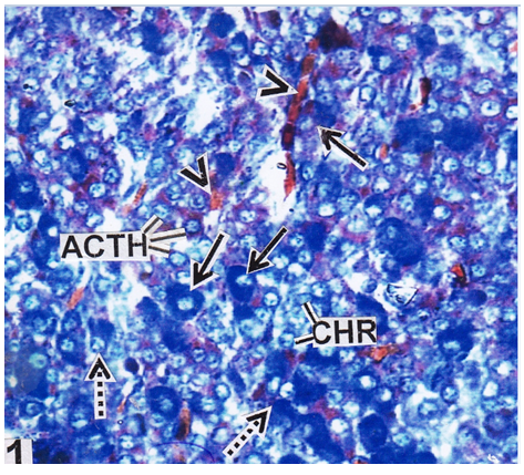
Fig: RPD region of pituitary gland during growth phase showing the distribution of GTH cells (solid arrows), TSH cells (broken arrows), ACTH cells and chromophobe cells (CHR). Note blood vessels (arrow heads) in between pituitary cells; (AB-OFG) × 400X.
Download Full Article: Click Here
Journal is Indexed and Abstracted in following Database(s).
    |
    |
       |